
Eyelid pathology
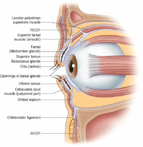
The eyelid is made up from the outside to the inside: skin, muscles allowing the eye to open and close, cartilage: the tarsus, eyelashes and conjunctiva.
Dr Bela, specialist in oculoplasty, treats all eyelid pathologies:
- upper eyelids:ptosis, dermatochalasis
- lower eyelids: ectropion, entropion
- eyelid tumors: benign (chalazion, sty, papilloma, nevus) and malignant (carcinoma).
The eyelids can be affected by different pathologies classified according to their etiology.
- These may be inflammatory diseases such as blepharitis which causes itching, loss of eyelashes, redness of the eyelids and dryness of the eye.
- These may be age-related degenerative diseases such as ptosis, ectropion or entropion. These latter pathologies lead to a drooping of the upper eyelid, by relaxation of the levator superior eyelid muscle on the eye, thus reducing its opening. Entropion and ectropion are degenerative pathologies of the lower eyelid linked to relaxation of the lower eyelid muscle. This causes the edge of the eyelid to roll inwards or outwards, causing the eyelashes to rub against the eye, chronic inflammation or daily tearing.
- Finally, these can be benign or malignant eyelid tumor pathologies. The most common benign tumors are chalazion, xanthelasma or cysts such as hydrocystoma for example. Among malignant tumors, the most common is basal cell carcinoma, but all types of skin tumors can occur in the eyelids.
All of these eyelid diseases most often require surgical intervention to restore the position of the eyelid or remove the tumor. When possible, particularly for inflammatory eyelid pathologies, conservative medical treatment will be offered.
Ectropion
Ectropion is a degenerative disease of the eyelid.
Following the overall relaxation of the lower eyelid muscle as well as the ligaments that hold the eyelid on either side of the eye, the lower eyelid lacks support. Following this loss of support for the muscle, tarsus and ligament complex, the eyelid will roll up on itself. When the free edge of the eyelid rolls outward, it is called ectropion. The eyelid is no longer in contact with the eyeball, the closure of the eye is incomplete and the lubrication of the eye is insufficient.
And this causes symptoms such as tearing, redness and irritation in the eyes. Furthermore, the edge of the eyelid is not well moistened. It will therefore become red and irritated and cause daily discomfort.
Different causes are possible:
- Most often it is an age-related pathology with a progressive loosening of the tarso ligamentous strap.
- Paralytic ectropion occurs as a result of facial paralysis
- Scarring ectropion occurs following a wound, a burn, or trauma to the skin of the lower eyelid which causes retraction of the eyelid. This will encourage the eyelid to roll outwards.
The only treatment for this eyelid pathology is surgery to tighten the eyelid ligament during this surgical procedure.
Here is some information about this surgery:
1 hour in outpatient surgery
local with sedation
- Anesthesia consultation before surgery
- Be fasting on the day of the operation
- Exit with a bandage that does not cover the eye
- Be accompanied when leaving
- Autonomy the same evening
Do not do sport, put your head under water or wear make-up for a week.
Swelling and bruising are systematic and disappear within 15 days.
Day 1: Appointment with Dr Bela to remove the dressing
Day 7: removal of sutures
M1: final result
Entropion
Entropion is a degenerative disease of the eyelid.
Following the overall relaxation of the lower eyelid muscle as well as the ligaments that hold the eyelid on either side of the eye, the lower eyelid lacks support. Following this loss of support for the muscle, tarsus and ligament complex, the eyelid will roll up on itself. When the free edge of the eyelid rolls inward, it is called entropion.
When the eyelid rolls toward the inside of the eye, against the eyeball. The free edge of the eyelid and the eyelashes of the eyelid will then rub against the surface of the eye. Symptoms are redness, pain and chronic irritation of the eye. This can have serious consequences on vision if not treated quickly with erosions of the surface of the eye and then scarring of the cornea impairing vision in the long term.
Different causes are possible:
- Most often it is an age-related pathology or senile entropion. Sometimes a spastic component is associated with a contraction of the orbicularis muscle in addition to the relaxation which maintains the invagination of the pulids.
- It can also be a consequence of pathologies of the conjunctiva inside the lower or upper eyelid. When the conjunctiva presents with inflammatory diseases, fibrosis or post-traumatic scarring, the eyelid will be drawn towards the inside of the eye and roll towards the eyeball.
This problem must be treated quickly to avoid consequences on the cornea and vision. The only treatment for entropion is eyelid surgery, which involves tightening the eyelid ligament and removing scar tissue if necessary. While waiting for surgery, the eye must be protected with ointment, a protective lens and sometimes an injection of botulinum toxin.
Here is some information about this surgery:
1 hour in outpatient surgery
local with sedation
- Anesthesia consultation before surgery
- Be fasting on the day of the operation
- Exit with a bandage that does not cover the eye
- Be accompanied when leaving
- Autonomy the same evening
Do not do sport, put your head under water or wear make-up for a week.
Swelling and bruising are systematic and disappear within 15 days.
Day 1: Appointment with Dr Bela to remove the dressing
Day 7: removal of sutures
M1: final result
Ptôsis
Ptosis is a disease of the levator upper eyelid muscles. There are two muscles that allow the upper eyelid to open:
- the levator muscle of the upper eyelid
- Muller’s muscle
When one of these two muscles is affected, the opening of the eye and the visual field of the eye is impaired. The eyelid droops over the eye, this is called ptosis.
Different causes are possible:
- Most often it is a degenerative disease linked to age with a relaxation of the levator palpebrae muscle which weakens and no longer opens the eye as effectively .
- In children, it may be a congenital ptosis present since birth.
- In adults ptosis can also be associated with neuromuscular, muscular (myopathy or myasthenia) or neurological (paralysis of a nerve of the third cranial pair) causes
- Finally it may be a false ptosis linked to a disease of the orbit called enophthalmia.
All these causes will be sought and eliminated in consultation in order to treat the underlying disease if necessary.
Ptosis requires surgery to raise the upper eyelid and allow sufficient and symmetrical opening of the eye on both sides.
The surgical technique depends on the type of ptosis and the age of the patient: plication reinsertion or resection of the levator palpe muscle, mullerian conjunctivo resection, frontal suspension.
Here is some information about this surgery:
1 hour in outpatient surgery
local with sedation
- Anesthesia consultation before surgery
- Be fasting on the day of the operation
- Exit with a bandage that does not cover the eye
- Be accompanied when leaving
- Autonomy the same evening
Do not do sport, put your head under water or wear make-up for a week.
Swelling and bruising are systematic and disappear within 15 days.
Day 1: Appointment with Dr Bela to remove the dressing
Day 7: removal of sutures
M1: final result
Benign or malignant eyelid tumor
Eyelid tumors are very common. They can be benign or malignant.
- The majority of eyelid tumors are benign.
They are safe and without any risk of spreading to other parts of the body.
These include, for example, chalazion, papilloma, nevus, or even cysts like hydrocystoma or deposits like xanthelasma. The treatment is either surgical or laser treatment can be carried out if no diagnosis is present.
- Malignant tumors of the eyelid are more difficult to treat because these pathologies systematically require surgical resection with time to reconstruct the upper or lower eyelid depending on the location.
Among the malignant eyelid tumors we can cite the most common: basal cell carcinoma. This one has a good prognosis. It presents a slow progression with very little risk of secondary locoregional or distant localization.
The treatment of eyelid tumors is only surgical and will depend on the nature of the tumor. For this, a biopsy must be carried out in the operating room to analyze the lesion. Once the diagnosis has been made, Doctor Bela will perform a tumor excision which must be complete.
Thanks to collaboration with a specialist anapathomopathologist present in the operating room, the tumor can be removed in its entirety after verification on site that the excision limits are healthy. Doctor Bela will then be able to reconstruct the eyelid so as not to leave any unsightly scars.
The more aggressive the tumor, the greater the resection margins must be and the more delicate the reconstruction is. Sometimes this surgical treatment must be combined with additional radiotherapy if the tumor is too large or at high risk of recurrence.
That’s why you should always seek medical advice as soon as you notice an eyelid tumour. Dr Bela will carry out a full examination of the eyelids at the surgery to determine whether the tumour is suspected of being malignant, and will quickly suggest the appropriate treatment.
The best way to prevent malignant eyelid tumours is to protect your skin on a daily basis with a sunblock, wear sunglasses and a hat, and avoid direct exposure to the sun. Lastly, it is advisable to schedule an annual screening consultation with your ophthalmologist, especially if you have a favourable background and a personal or family history of skin tumours.
Here is some information about this surgery:
30 minutes to 2 hours depending on the size of the tumour, in outpatient surgery or with overnight hospitalisation
local with sedation or general anaesthesia
- Anesthesia consultation before surgery
- Be fasting on the day of the operation
- Exit the same day or next day with a dressing that covers the eye for 24 hours
- Be accompanied when leaving
Do not do sport, put your head under water or wear make-up for a week.
Swelling and bruising are systematic and disappear within 15 days.
Day 1: Appointment with Dr Bela to remove the dressing
D7 or D15: removal of sutures depending on the surgical reconstruction
M1: final result
What are eyelid diseases?
There are several types of disease that can affect the upper or lower eyelids. Benign diseases are inflammatory diseases such as blepharitis, chalazion or stye. There may also be diseases of the eyelid conjunctiva, such as allergic conjunctivitis or infectious bacterial or viral conjunctivitis. Degenerative pathologies such as ptosis, entropion or ectropion may also be involved. The most serious diseases are malignant eyelid tumours, which must be detected and treated quickly to prevent their spread.
What are the symptoms of blepharitis?
Blepharitis is linked to an engorgement of the glands located inside the upper and lower eyelids. These glands, known as the Meibomian glands, are responsible for secreting fat into the tears. The purpose of this secretion is to protect the tears from evaporation and thus ensure that the surface of the eye is always hydrated. When these glands become blocked, the fatty layer in the tears is reduced, leading to dry eyes and reflex watering. The eyelids also become inflamed and red, which causes itching and can eventually lead to the loss of eyelashes.
What is blepharospasm?
Blepharospasm is a pathology affecting the muscles around the eyes: the eyelid muscles and more specifically the orbicularis muscle. The orbicularis muscle is responsible for the blinking and forced closure of the eyelids. It is innervated by a nerve called the facial nerve, which originates in the brain and has a voluntary contraction. Forced closure of the eye is therefore normally a voluntary process in the brain. Blepharospasm is a condition that affects this facial nerve, causing involuntary stimulation. This causes the eye to close involuntarily and uncontrollably. This condition is very disabling in everyday life, as it can lead to falls. It may be involuntary or associated with a neurological malformation, but in most cases no aetiology is found. The only effective treatment is a lifelong injection of botulinum toxin into the muscle responsible for the disease.
Can droopy eyelids be lifted without surgery?
It is possible to lift drooping eyelids without surgery, thanks to cosmetic medicine procedures. Botulinum toxin is used to reduce wrinkles around the eyes and can also be used to raise the eyebrows to open up the eyes. Hyaluronic acid is used to fill in the hollows under the eyes and make dark circles disappear, but it can also be used to support the eyebrow, raising it and opening up the eyes. Certain aesthetic medicine techniques can be used, such as lasers, to reduce excess skin on the upper eyelid. In some cases, surgery is essential to lift drooping eyelids. This involves removing the excess skin known as dermatochalasis. This will be discussed in consultation with your surgeon.
How can I tell if an eyelid tumour is benign or malignant?
There are certain criteria for assessing whether a recent lesion or pimple on the eyelid is a benign or malignant tumour. The first step is to assess the patient's background. Certain conditions are more favourable, such as a family or personal history of skin tumours, genetic diseases that predispose to tumours, the type of skin known as the phototype, sun exposure, smoking and a history of radiotherapy. In the case of an eyelid tumour, the aim is to determine whether it has developed rapidly and aggressively, leading to suspicion of malignant disease. More often than not, malignant eyelid tumours are also associated with bleeding, itching, surface excoriations and induration to the touch, with the tumour appearing to be attached to deep tissue. Finally, a loss of eyelashes are all elements that raise the suspicion of a malignant tumour. The only way to be sure of the diagnosis is to carry out surgery to remove some or all of the tissue so that it can be analysed under a microscope.